- What is Computed Radiography?
- Components of Computed Radiography
- How Does Computed Radiography Work?
- How is CR Different from DR?
- Applications of Computed Radiography
In the fast-changing world of medical imaging, Computed Radiography (CR) systems have emerged as a cornerstone technology, bridging the gap between traditional film-based radiography and modern digital imaging solutions. This blog aims to share a comprehensive overview of CR systems, their functionality, advantages, and their crucial role in the medical imaging landscape.
What is a CR System?
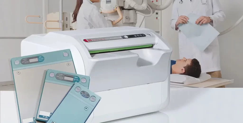
Computed Radiography (CR) is a digital imaging technology that utilizes phosphor imaging plates to capture X-ray images. Unlike conventional film radiography, which requires chemical processing to develop images, CR systems convert X-rays into digital signals, enabling quick and efficient image processing, storage, and sharing.
Components of Computed Radiography
Computed Radiography (CR) systems comprise several key components that capture, process, and display digital X-ray images. Understanding these components is crucial for appreciating how CR systems function and their role in medical imaging. Here are the main components of a CR system:
1. X-ray Source
• X-ray Tube: This tube generates X-rays that pass through the patient’s body to create an image. It is a crucial part of any radiography system, including CR.
2. Imaging Plate (IP)
• Phosphor Plate: The core component of a CR system is the imaging plate, which is coated with photostimulable phosphor material that captures and stores X-ray energy as a latent image.
3. CR Reader/Digitizer
• Plate Transport Mechanism: Moves the imaging plate into the reader for processing.
• Laser Scanner: A laser beam scans the imaging plate, releasing the stored energy as visible light.
• Photodetector: Captures the emitted light from the imaging plate and converts it into an electrical signal.
• An analog-to-digital converter (ADC) converts the electrical signals from the photodetector into digital data, forming the digital image.
4. Image Processing Software
• Processing Algorithms: These algorithms enhance the digital image by adjusting contrast, brightness, and sharpness to improve diagnostic quality.
• Quality Control Tools: Ensure the image meets specific diagnostic criteria and standards.
5. Image Display System
• Monitor: High-resolution display monitors allow radiologists to view and analyze digital images. Monitors designed for medical imaging typically offer superior resolution and grayscale capabilities compared to standard monitors.
6. Image Storage and Management
• Picture Archiving and Communication System (PACS): A digital storage system for managing and retrieving medical images, PACS allows images to be stored, accessed, and shared efficiently across healthcare facilities.
• Database Management System: Organizes and indexes the digital images within the PACS for easy retrieval.
7. Workstation
• Computer: Processes and displays digital images, often equipped with specialized image enhancement and analysis software.
• Input Devices: Keyboards, mice, and sometimes specialized tools for navigating and manipulating images.
8. Networking Infrastructure
• Network Connectivity: Facilitates the transfer of digital images between the CR system, PACS, and other hospital information systems (HIS/RIS).
9. Backup and Archiving Systems
• Data Backup Solutions: These solutions ensure the safety and redundancy of stored images, preventing data loss in case of hardware failure.
• Archiving Solutions: Long-term storage solutions, such as cloud or offsite servers, maintain historical image records.
Additional Accessories
• Cassette: Houses the imaging plate and protects it from light and physical damage during handling.
• Barcode Reader: This device is often used to link patient information with the imaging plate, ensuring accurate identification and record-keeping.
Workflow Integration
• Radiology Information System (RIS): This integrates with the CR system to manage patient data, imaging orders, and scheduling, enhancing overall workflow efficiency.
By understanding these components, healthcare providers can appreciate how CR systems streamline capturing, processing, and managing digital X-ray images, ultimately enhancing diagnostic capabilities and patient care.
How Does a CR System Work?
1. Image Acquisition:
• X-ray Exposure: The patient is exposed to X-rays captured by a phosphor imaging plate instead of traditional film.
• Imaging Plate: The phosphor plate stores the X-ray energy as a latent image.
2. Image Processing:
• Plate Scanning: A CR reader or digitizer then scans the imaging plate. The plate is exposed to a laser, which releases the stored X-ray energy as light.
• Digital Conversion: The light emitted by the plate is captured by a photodetector and converted into digital signals. These signals are processed to create a digital image.
3. Image Review and Storage:
• Image Display: The digital image is displayed on a computer monitor for immediate review by radiologists and clinicians.
• Image Storage: The images can be stored in Picture Archiving and Communication Systems (PACS) for easy retrieval and long-term storage.
Applications of CR Systems
1. General Radiography:
• CR systems are widely used in routine radiographic examinations, including chest X-rays, skeletal imaging, and abdominal imaging.
2. Emergency and Trauma:
• The speed and efficiency of CR make it ideal for emergency and trauma settings where quick diagnostics are critical.
3. Pediatric Imaging:
• Reduced radiation doses make CR systems suitable for pediatric patients, ensuring safety while maintaining image quality.
4. Mammography:
• Advanced CR systems are also used in mammography, providing detailed breast imaging for early detection of abnormalities.
CR VS DR
Computed Radiography (CR) uses a cassette-based system to capture X-ray images. The X-ray cassette contains a photostimulable phosphor plate, which captures the image when exposed to X-rays. The cassette is then inserted into a CR reader, where a laser scans the plate to convert the latent image into a digital format. This digital image can then be viewed, enhanced, and stored electronically.
Advantages:
• Cost-Effective: CR systems are generally less expensive than DR systems, making them a good option for facilities with budget constraints.
• Compatibility: CR systems can be used with existing X-ray equipment, allowing an easier transition from traditional film-based radiography.
• Flexibility: CR plates come in various sizes, accommodating different imaging needs and body parts.
Disadvantages:
• Processing Time: The need to transfer cassettes to a reader adds an extra step, making CR slower than DR.
• Image Quality: While good, the image quality of CR is typically lower than that of DR.
Digital Radiography (DR)
Digital Radiography (DR) uses flat-panel detectors to capture X-ray images and convert them into digital signals directly. These detectors can be fixed or portable and integrated with the X-ray system. The digital images are available almost instantaneously on a computer screen for viewing, enhancement, and storage.
Advantages:
• Speed: DR systems provide immediate image availability, significantly reducing the time between exposure and diagnosis.
• Image Quality: DR generally offers superior image quality with higher resolution and better contrast, aiding in more accurate diagnoses.
• Efficiency: The workflow is streamlined, as there is no need to handle cassettes, making DR ideal for high-volume imaging environments.
• Lower Radiation Dose: DR systems often require lower radiation doses to produce high-quality images than CR.
Disadvantages:
• Cost: DR systems are more expensive upfront than CR systems, which can be a barrier for smaller facilities.
Conclusion
Choosing between Computed Radiography (CR) and Digital Radiography (DR) depends on your medical imaging facility’s specific needs and circumstances. CR offers a cost-effective solution that is compatible with existing X-ray equipment, making it suitable for facilities with budget constraints or those transitioning from traditional film. However, the additional processing step and slightly lower image quality may be limitations.
On the other hand, DR provides immediate image availability, superior image quality, and a streamlined workflow, making it ideal for high-volume environments and facilities, prioritizing efficiency and diagnostic accuracy. The higher upfront cost of DR can be offset by its long-term benefits, including reduced processing time and potentially lower radiation doses.
Many factors should be taken when you need to buy x-ray machines, such as prices, volume of imaging, desired image quality, and workflow efficiency. Both CR and DR have unique advantages, and selecting the right system will enhance the overall effectiveness of your imaging services.


