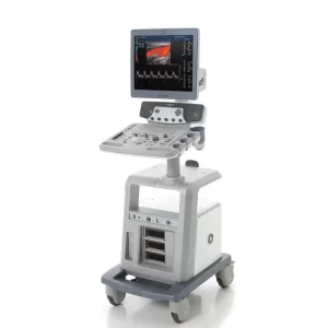
Features
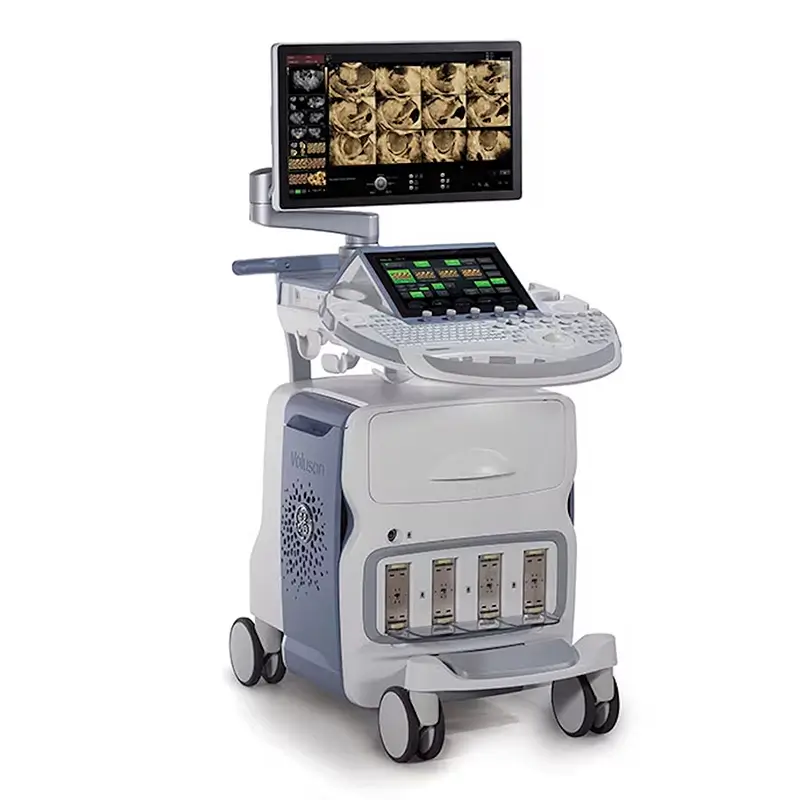
- Up to 40 frames per second in 4D mode
- 2D/3D/4D Imaging
- “HDlive” advanced 4D ultrasound imaging
- 67,584 imaging channels
- M-Mode, M-Color Flow
- PW/CW Spectral Doppler
- Color/Power/Tissue Doppler
- HD Flow
- B-Flow
- CrossXBeam
- SRI II HD Speckle Reduction Imaging
- Coded Excitation
- Coded Harmonics
- Contrast Imaging
| Dimensions | 50.8”,1290mm (h) x 22.8”, 580mm (w) x 36.6”, 930mm (d) |
| Weight | 265lbs,120kg |
| Monitor | 15” LCD Monitor High brightness with 350 cd/m2 Tilt/Rotate adjustable Monitor Tilt Angle: +10°/-90° Rotate Angle: 360° Digital brightness & contrast adjustment |
| Console Design | Digital brightness & contrast adjustment 3 Active Probe ports ( plus 1 non-imaging port) Integrated HD (160 GB) Integrated DVD+ R(W) / CD-R(W) drive On-board storage for peripherals Wheels – Wheel diameter 150mm Integrated locking mechanism that provides rolling lock Integrated cable management Front and rear handles |
| Operator Keyboard | Floating Keyboard: Rotation: adjustable+/-40° from center Height adjustable + 200mm Full-sized, backlit alphanumeric keyboard Ergonomic hard key layout Interactive back-lighting Intergrated recording keys for remote control of up to 4 Peripherals or DICOM devices |
| Touch Screen | 10.4 in High-Resolution color CD screen Interactive dyamic software menu Brightness adjustable |
| System Standard | State-of-the-art user interface with high resolution 10.4inch LCD touch panel Automatic Tissue Optimization Tissue doppler Coded Harmonic Imaging Coded Excitation (CE) HD-Flow XTD SRI III (Speckle reduction Imaging) CrossXBeamCRI (Compound Resolution Imaging) Focus&Frequency composite (FFC) High resolution Zoom Pan Zoom Steering Virtual Convex Beta-View Patient Information Database Image Archive on hard drive 3D/4D data compression (lossy/Lossless) Inversion Real-time automatic Doppler calcs Measurement & Calculations including Worksheets/Reports for: OB, GYN, Vascular, Cardio, Abdominal, Small-Parts, Urology, Pediatrics, Ortho, Neurology Mutigestational calculations |
| Operation Mode | B-Mode (2D) M-Mode (M) M-Color-Mode (MC) Color Flow Mode (C) Power Doppler Imaging (PD) Tissue Doppler Imaging(TD) HD-Flow Imaging (HD-Flow) PW Doppler with high PRF (PW) B-Flow Extended View (XTD View) Coded Contract Imaging (Contrast Media) Volume Mode(3D/4D): – 3D static – 4D Real Time – VCI-A, VCI-C – STIC/ Color, Angio, HD-Flow, Contrast & B-Flow – 4 D Biopsy |

