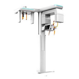
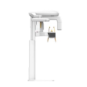
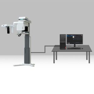
CBCT
What is Cone-Beam CT and How Does it Work?
Cone-beam CT is a specialized imaging technology that provides detailed three-dimensional images of the teeth, jawbone, and surrounding structures.
Cone-beam CT principle is that the X-ray generator performs a circular digital projection around the projection body. After the rays pass through the photographed object, they are received by the flat-panel detector. X-rays are converted into electrical signals and sent to the computer for image reconstruction to obtain a three-dimensional image.
What are the Core Components of CBCT?
CBCT consists of CT, panoramic flat-panel detector, lateral flat-panel detector, and focus tube. Here are the benefits of the core components:
(1) CT Tube: The Toshiba tube is lighter than domestic tubes and has a smaller rotating armload, more stable operation, and clearer imaging.
(2) Flat-panel detector: large field of view, stable performance and longevity, and high imaging quality
What is the Difference CBCT and Traditional CT Scan?
CBCT and traditional CT scanners are both imaging techniques that use X-rays to produce detailed cross-sectional images of the body. Here are the main distinctions:
(1) CBCT provides continuous imaging throughout the scan. Traditional CT produces a single slice image per scan.
(2) Conventional CT scan has a larger field of view than CBCT
(3) CBCT generally has a lower radiation dose than traditional CT scans.
Showing all 3 resultsSorted by average rating
-
CBCT
Dental Cone-Beam Computed Tomography UzzDFT-4D-COMMANDER(A)
Price range: $45,380.00 through $48,800.00


