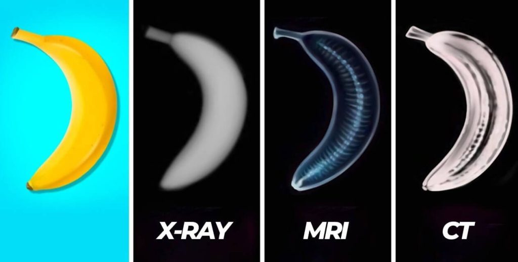
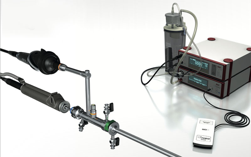
In medical diagnostics, imaging technologies such as MRI, CT scanners, and X-ray machines play a pivotal role. Each has unique characteristics, advantages, and specific use cases. Understanding the differences between MRI, CT scanners, and X-ray machines can help patients and healthcare providers make informed decisions about diagnostic options.
MRI (Magnetic Resonance Imaging)
Magnetic Resonance Imaging (MRI) utilizes strong magnetic fields and radio waves to create detailed images of the body’s internal structures. The process involves hydrogen atoms in the body’s tissues resonating in response to the magnetic field. A computer then detects and processes these resonant signals to produce high-resolution images.
Uses: MRI is efficient for imaging soft tissues, making it invaluable for examining the brain, spinal cord, joints, muscles, and internal organs. It is often used to diagnose neurological disorders, musculoskeletal conditions, cardiovascular diseases, and cancers.

Advantages:
- No Ionizing Radiation: MRI does not use ionizing radiation, making it safer for repeated use.
- High-Contrast Images: It provides excellent contrast between different types of soft tissues.
- Multi-Planar Imaging: MRI can produce images in multiple planes (axial, sagittal, and coronal) without moving the patient.
Disadvantages:
- Expensive: MRI machines and scans are typically more costly than other imaging modalities.
- Longer Exam Times: MRI scans can take longer, sometimes up to an hour, depending on the imaged area.
- Not Suitable for All Patients: Due to the strong magnetic fields, patients with metal implants or devices (like pacemakers) may be unable to undergo MRI.
CT Scanner (Computed Tomography)
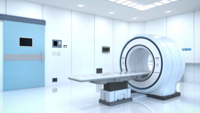
Principle: Computed Tomography (CT) uses X-rays to create cross-sectional images of the body. The X-ray tube rotates around the patient during a CT scan, capturing multiple photos from different angles. A computer then processes these images to create detailed cross-sectional views and three-dimensional reconstructions.
Uses: CT scans are widely used for various diagnostic purposes, including detecting tumors, internal injuries, infections, and vascular diseases. They are instrumental in emergencies where quick, detailed imaging is required.
Advantages:
- Detailed Cross-Sectional Images: CT scans provide highly detailed images of internal structures.
- Fast Scanning: The scanning process is quick and often completed within minutes.
- Versatile: CT scans can effectively image bones, soft tissues, and blood vessels.
Disadvantages:
- Ionizing Radiation: CT scans involve exposure to higher doses of ionizing radiation than standard X-rays.
- Cost: While typically less expensive than MRI, CT scans are still relatively costly.
- Soft Tissue Contrast: CT scans generally provide lower contrast between different types of soft tissues compared to MRI.
X-Ray (Radiography)
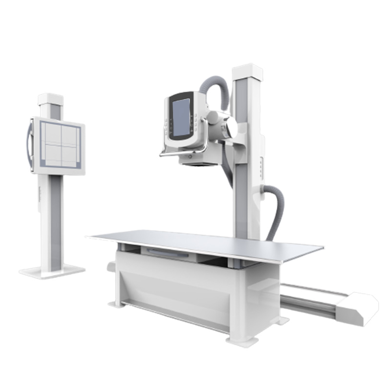
Principle: X-ray imaging uses high-energy electromagnetic waves to make images of the body inside. Different tissues absorb X-rays to varying degrees, with dense structures like bones absorbing more X-rays and appearing white on the X-ray film, while less dense tissues appear in shades of gray.
Uses: Mainly for diagnosing fractures, joint dislocations, chest conditions (such as pneumonia or lung cancer), and dental examinations. They are also used in mammography for breast cancer screening.
Advantages:
- Quick and Accessible: X-ray exams are fast and widely available.
- Low Cost: X-ray imaging is generally less expensive than MRI and CT scans.
- Effective for Bones: X-rays provide excellent images of bones and are highly effective for detecting fractures and other bone-related conditions.
Disadvantages:
- Ionizing Radiation: X-rays involve exposure to ionizing radiation, though typically in lower doses than CT scans.
- Limited Soft Tissue Detail: X-rays are less effective at providing detailed images of soft tissues.
- Two-Dimensional Images: X-ray images are two-dimensional and may not offer comprehensive views of complex structures.
Choosing the Right Imaging Modality
The choice between MRI, CT, and X-ray imaging depends on various factors, such as your health condition, the area of the body being examined, and the need for detail and clarity in the images. Here are some general guidelines:
- MRI is suitable for soft tissue imaging, with no radiation and longer imaging times.
- CT Scans are ideal for rapid, detailed imaging of internal organs, bones, and blood vessels, making them valuable in emergencies and for comprehensive diagnostic assessments.
- X-rays are most effective for quick evaluations of bone fractures, joint dislocations, and chest conditions, providing a cost-effective and widely accessible imaging option.
MRI, CT scans, and X-rays are all indispensable tools in modern medicine. Their unique attributes and applications ensure that healthcare providers can offer precise and effective diagnostic services, ultimately leading to better patient outcomes. Understanding when and how to use each modality is critical to leveraging their full potential in medical practice.

