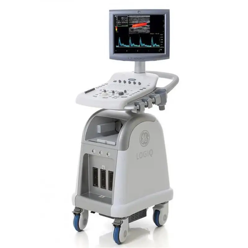
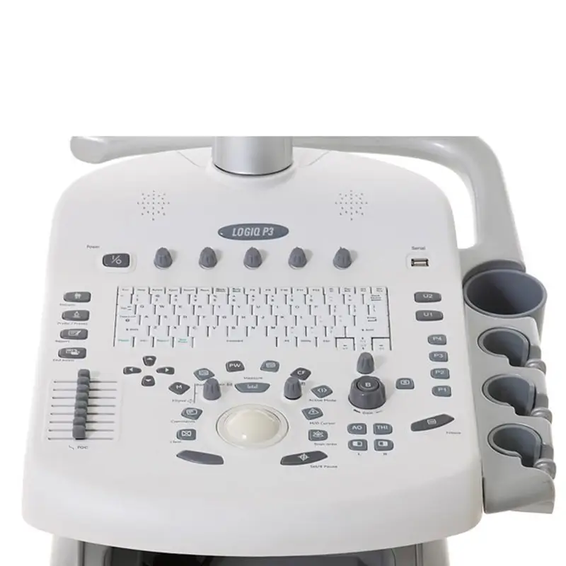
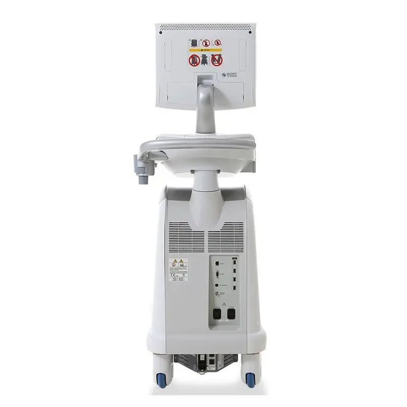
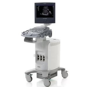
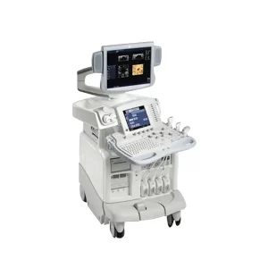
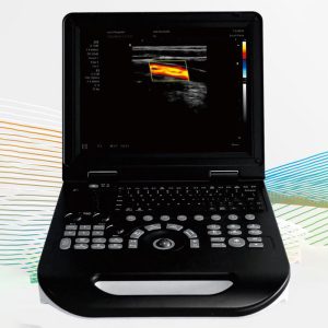
The GE Logiq P3 is an ultrasound system that offers color Doppler images. The Logiq P3 provides imaging capabilities for a variety of applications including; Cardiology, Gastroenterology, Neurology, Gynecology, and more. The GE Ultrasound system offers a 15-inch LCD monitor on a swivel arm, making it easy for both the operator and the patient to view the images. The System offers b-mode, m-mode, color flow Doppler, and power Doppler along with other imaging modes. With Imaging technologies like Cross X-beam, Tissue Harmonics, and Speckle reduction imaging you can get clear ultrasound images.
Features
- 2D, M-Mode
- Color/Power/PW Doppler
- Anatomic M-Mode
- Auto Optimization one-touch image enhancement
- 2 probe ports
- 15″ LCD screen
- DICOM
- LogiqView panoramic imaging option
- On-Board DVD-RW
- USB port for export
- Ethernet port for network connectivity
Technical Parameter
| Monitor | 15 inch High-Resolution Color LCD Display size: 1024 x 768 Interactive Dynamic Software Menu Open Angle Adjustab le, – 0 to 160° Brightness and Contrast Adjustment |
| Title/rotate adjustable monitor | Yes |
| Touchscreen | No |
| Trackball or Trackpad | Trackball |
| CP Backlight | Yes |
| Battery | No |
| Maximum depth of field | 30 cm |
| Minimum depth of field | 0-2 cm |
| Cart (HCU) | No |
| Independent steer & lockable wheels | Yes |
| Console Design | Integrated HOD (160 GB) 3 Probe Ports I 2 Probe Ports Rear Handle Wired LAN Support User Interface |
| Operator Keyboard | Alphanumeric Keyboard Ergonomic Hard Key Operations Integrated Recording Keys for Remote Control of Peripheral Devices and DICOM Devices 8 TGC Pods, with Re-mapping. Functionality at any Depth |
| Electrical Power | Voltage:100-120 Vac or 220-240 Vac Frequency:50/60 Hz POWER:Maximum 425 VA with Peripherals |

