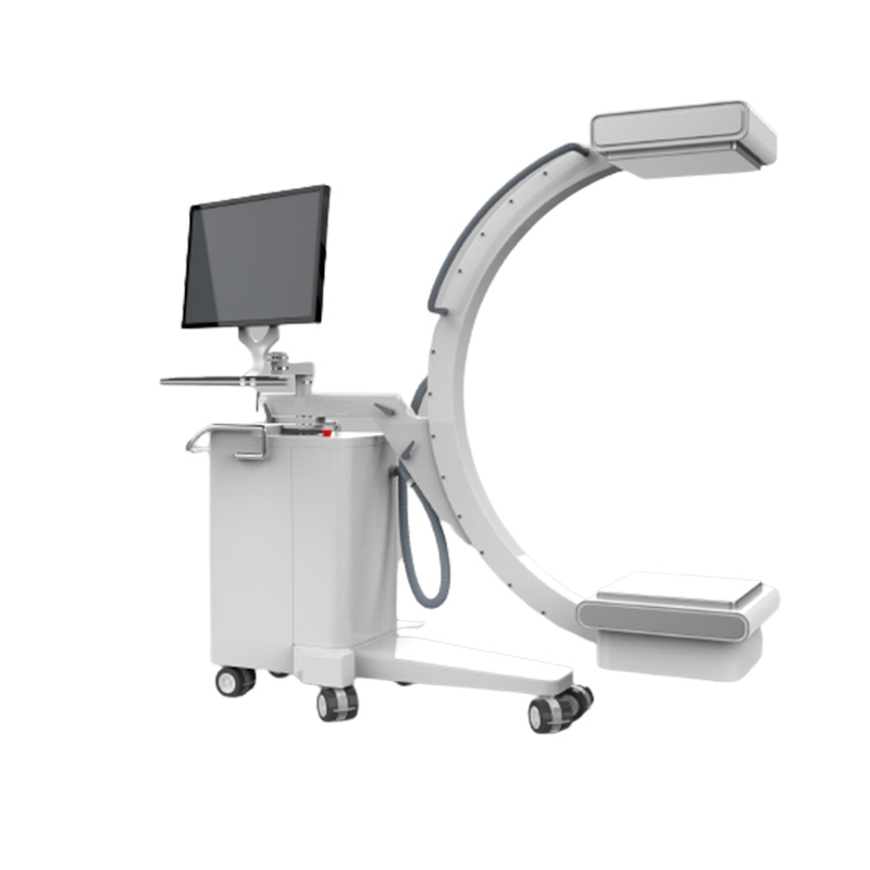
C arm machine features
- Precise full balance design allows the C-arm can be freely locked at any position, for easy operation and safe use
- Large opening and high-strength C-arm design can provide greater operating space for your operation
- Professional tube design for flat C-arm to provide enough space for your position
- Split, integrated design, handheld iPad, a variety of modes to choose from, free to provide convenient services for your use
1. Flat Panel Detector
- Type of the detector: TFT monolithic amorphous silicon
- Panel size of the detector: 210mm*210mm
- Acquisition pixel matrix of the detector: 1024×1024
- The pixel pitch of the detector: 200 um
- The spatial resolution of the detector: 2.5LP/MM
- Pixel grayscale: 16-bit
- Acquisition frame frequency: 30fps
2. High frequency and high voltage generator
- Max. Output power: 5kw
- Input power supply: 220 VAC
- Photography voltage: 40~125kV
- Photography tube current range: 10~100mA
- Exposure time range: 1ms-5000ms
- mAs range: 0.1mAs~200mAs
- Fluoroscopy voltage: 40~120kV
- Continuous fluoroscopy mA:0.5-5mA
- Pulse fluoroscopy mA:10-30mA
- Pulse fluoroscopy frame frequency: 1-30 frame
- Diagnostic self-test and display
3. X-ray bulb tube assembly
- Focus: 0.3mm/0.6mm
- Max.KV: 125kV
- Anode target angle: 12°
4. C arm stand
- Vertical displacement of C arm (electric): ≥400mm
- Horizontal displacement of C arm (manual): ≥200mm
- Rotation displacement of C arm (manual): ≥±200°
- Horizontal hunting displacement of C arm (manual): ≥±12.5°
- Track displacement of C arm (manual): -30°±5°~+90°±5°
- Depth of arc of C arm ≥650mm
- S.I.D:≥1000mm
5. The grid and the beam-limiting device
- The grid material: Aluminum-based grid
- Size: 23×23cm
- Grid ratio: 100:1
- Wire/inch: 215C
- Grid Focal length: 1000mm
- Electric C arm DR beam limiting device
6. Workstation functions for image acquisition, processing, and diagnosis
- The Chinese user interface, standard DICOM3.0 image
- Workstation functions for image acquisition: adjust or preset window width/ window level, local automatic window level, preset window width/ window level, positive and negative image flipping, image flipping, rotation, image magnification and roam, image interpolation edge enhancement, local magnification, restoration, image annotation, text annotation/number annotation, image marking, ruler line segment measurement, square and round measurement, arbitrary shape measurement, angle measurement, automatic electron sharing, image stitching, acquisition and display of exposure index
- Software package for the special image acquisition control and software package for special parts protocol processing
- Functions of patient management, image acquisition, image processing (image correction, image flipping, USM sharpening, image filtering), image observation (provide the tools for observation and measurement)
- Image printing, DICOM printing, paper printing, manual printing for the displayed images, a single button for marking and printing the images, optional for different printers, film format, and number of prints, print queue control, stop/start the preset
- User personalization: showing the format and layout, default setting, toolbar setting, and parts protocol enhancement filter
- Image display: display configuration 1920×1080, HD display
7. Workstation
- CPU: Intel i7,3GHz or above
- Host memory: ≥16GB DDR3 1600 high speed memory
- Hard disk: 1T/7200rpm large capacity and high-speed hard disk
- Workstation monitor: IPS LCD monitor
- Network card and network interface: 1000M network card, 1000M network interface, Jumbo Frames: 9K
- DICOM3.0 interface
- Windows 10 64-bit SP1 (Professional Edition or higher)


